
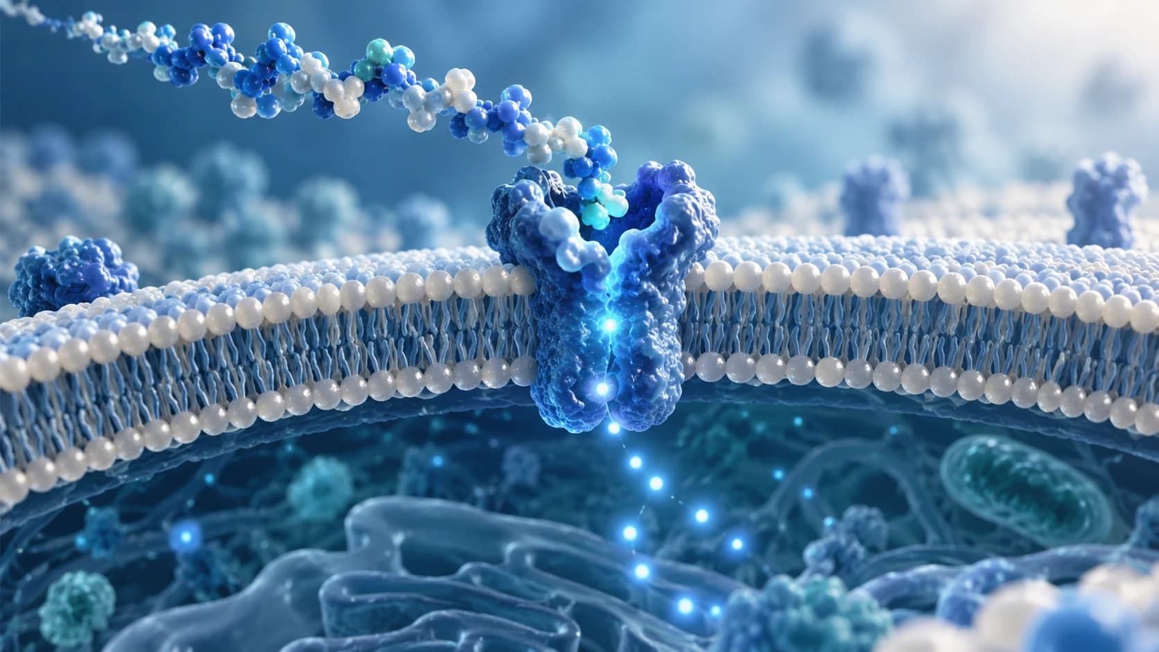
This article is for general education and research information. It does not provide medical advice or instructions for personal use.
Key Takeaways
- Peptide signalling begins when a peptide reaches a responsive cell and interacts with a compatible receptor.
- Many peptide hormones communicate through cell-surface receptors, which transfer an extracellular message into the cell.
- Different receptors activate different pathways. GLP-1 receptors primarily signal through G proteins and cAMP, while insulin uses a receptor tyrosine kinase.
- Second messengers such as cAMP, IP3 and calcium carry receptor-generated signals to targets inside the cell.
- The same peptide can produce different responses in different tissues because receptor expression and intracellular signalling machinery vary.
- Cells limit peptide signals through peptide breakdown, second-messenger removal, receptor desensitisation and receptor trafficking.
- Showing that a peptide activates a receptor or pathway does not by itself prove a meaningful health benefit in humans.
Peptide signalling is the process by which certain peptides carry biological messages between cells. A signalling peptide reaches a responsive cell, interacts with a receptor and triggers changes inside that cell.
Insulin provides a familiar example. Pancreatic beta cells release insulin when blood glucose rises. The hormone enters the circulation and reaches many tissues, but a cellular response depends on the presence of an insulin receptor and the signalling machinery connected to it.
Other peptide signals work over much shorter distances. Some are released by nearby cells, while neuropeptides participate in communication within the nervous system.
The basic principle remains the same: a peptide message is released, recognised and converted into a cellular response.
This process is part of the body's larger cell signalling system. If you first need the molecular basics, read What Peptides Are and How They Work.
What Peptide Signalling Means
Some peptides function as ligands, molecules that interact with particular receptors.
A useful overview of peptide signal transduction is:
Peptide release → transport → receptor binding → signal transduction → cellular response → signal termination
| Stage | What happens |
|---|---|
| Release | A cell secretes a signalling peptide |
| Transport | The peptide moves locally or through the circulation |
| Reception | The peptide interacts with a compatible receptor |
| Transduction | Receptor activation starts signalling inside the cell |
| Response | Cellular activity changes |
| Termination | The signal is reduced, redirected or switched off |
Signal transduction is the conversion of an extracellular message into intracellular biochemical activity.
That distinction is useful because simply detecting a peptide in blood or tissue does not show what effect it will have. The receiving cell must be able to recognise the signal and translate it into a response.
Depending on the receptor and cell type, that response can affect enzyme activity, secretion, ion movement, metabolism, transport processes or gene expression.
How Peptide Messages Reach Their Target Cells
Peptide messages do not all travel the same distance.
- In endocrine signalling, a hormone enters the circulation and reaches responsive cells in other tissues. Insulin is one example.
- In paracrine signalling, a released molecule acts mainly on nearby cells in the surrounding tissue.
- In autocrine signalling, a cell can respond to a signal that it releases itself.
- Neuroendocrine signalling occurs when specialised neurons release signalling molecules into the circulation, allowing neural activity to influence distant tissues.
These terms describe how the message reaches its target. They do not tell us which intracellular pathway will be activated after receptor binding.
That next step depends on the receptor.
Why Many Peptide Hormones Use Cell-Surface Receptors
A human cell is surrounded by a plasma membrane that separates its interior from the extracellular environment.
Many familiar peptide hormones act through receptors embedded in this membrane. GLP-1, for example, acts through the GLP-1 receptor, while insulin acts through the membrane-spanning insulin receptor. The IUPHAR/BPS Guide to Pharmacology identifies the GLP-1 receptor as a G protein-coupled receptor, while its insulin receptor family entry classifies the insulin receptor as a receptor tyrosine kinase.
These receptors allow an extracellular peptide signal to change events inside the cell without requiring the peptide itself to carry the message directly through the membrane.
A receptor is not simply a docking point. Ligand binding can change receptor activity or conformation, and that change starts the biochemical events responsible for peptide receptor signalling.
Being a peptide also does not automatically predict how a molecule behaves. Molecular size and structure should be separated from biological function, a distinction explained further in Peptides vs Proteins.
How GPCR Signalling Carries a Peptide Message Into the Cell
One major receptor family involved in peptide signalling is the G protein-coupled receptor, or GPCR.
GPCRs connect ligand binding outside the cell with G proteins on the intracellular side of the membrane.
The GLP-1 receptor provides a well-characterised example. The IUPHAR/BPS Guide to Pharmacology lists the Gs family and stimulation of adenylyl cyclase as its primary transduction mechanism.
A simplified GLP-1 signalling sequence is:
GLP-1 → GLP-1 receptor → Gs → adenylyl cyclase → cAMP → downstream response
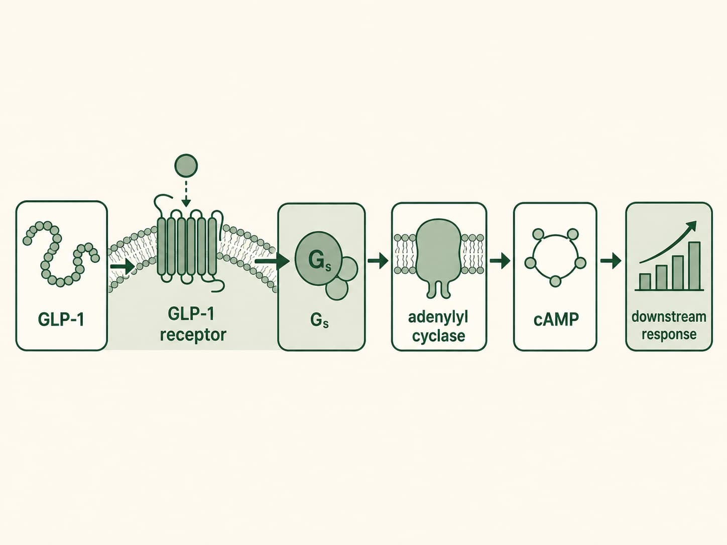
Figure: GLP-1 signalling pathway. GLP-1 binds to the GLP-1 receptor, activating the Gs protein and adenylyl cyclase, which increases cAMP signalling and triggers downstream cellular responses.
After GLP-1 activates its receptor, the receptor can activate a G protein. Gs then stimulates adenylyl cyclase, an enzyme involved in producing cyclic adenosine monophosphate, better known as cAMP.
cAMP is a second messenger. The peptide is the extracellular message, while cAMP helps carry information from the activated receptor to proteins elsewhere inside the cell.
Research in pancreatic beta cells has directly measured changes in cAMP associated with GLP-1 receptor activity, showing how receptor signalling can be connected to changes in intracellular messenger levels. A 2021 study using fluorescent cAMP measurements in mouse and human beta cells examined this relationship in detail.
But GPCR does not automatically mean cAMP.
Vasopressin demonstrates another route. Human vasopressin interacts with several receptor subtypes, including V1A, V1B and V2. The IUPHAR/BPS receptor database identifies Gq/G11 as the principal transduction family for the V1A receptor.
That pathway can be simplified as:
Vasopressin → V1A receptor → Gq/G11 → phospholipase C → IP3 and DAG → calcium-related signalling
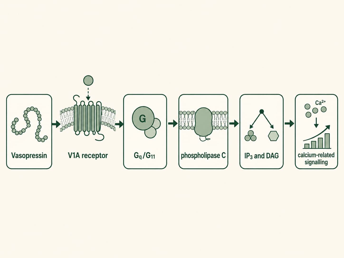
Figure 2: Vasopressin V1A receptor signalling pathway. Vasopressin binds to the V1A receptor and activates Gq/G11, which stimulates phospholipase C to generate IP3 and DAG, leading to increased intracellular calcium signalling and downstream cellular responses.
The IUPHAR/BPS V1A receptor entry specifically identifies phospholipase C stimulation as a primary response downstream of Gq/G11.
This is why there is no universal “GPCR peptide pathway.” Receptor subtype determines which signalling machinery is recruited.
Why Insulin Uses a Different Signalling Route
Peptide signalling is not synonymous with GPCR signalling.
Insulin uses a different receptor system.
The IUPHAR/BPS Guide to Pharmacology places the insulin receptor in the Type II receptor tyrosine kinase family. After receptor activation, signalling includes rapid autophosphorylation of intracellular tyrosine residues followed by recruitment of adaptor proteins such as IRS1 and IRS2.
A simplified sequence is:
Insulin → insulin receptor → tyrosine phosphorylation → IRS proteins → downstream signalling
This contrast with GLP-1 makes an important point:
| Feature | GLP-1 | Insulin |
|---|---|---|
| Receptor family | GPCR | Receptor tyrosine kinase |
| Early signalling event | G-protein activation | Tyrosine phosphorylation |
| Key downstream components | Adenylyl cyclase and cAMP | IRS proteins and kinase networks |
| Final response | Depends on target cell | Depends on target cell |
Both are peptide hormones, but they do not use the same intracellular signalling pathway.
That is why classifying molecules simply as “peptides” tells us relatively little about their biological effects.
How Second Messengers Amplify And Shape Peptide Signals
Receptor binding starts the message. Intracellular signalling determines how that message is distributed through the cell.
Second messengers such as cAMP, IP3, DAG and calcium ions can connect an activated receptor to multiple downstream proteins.
The cAMP pathway gives a useful example. Activation of a Gs-coupled receptor can stimulate adenylyl cyclase, increasing intracellular cAMP. cAMP can then regulate downstream effectors rather than limiting the response to the receptor itself. Experimental work in beta cells confirms that changes in cAMP can be closely linked to GLP-1 receptor signalling.
This type of cascade allows a signal to be amplified: an event at the receptor can influence an enzyme, which can generate intracellular messengers that act on additional targets.
The result is not a fixed multiplier shared by every receptor. Signal strength depends on receptor number, ligand concentration, downstream enzymes, messenger breakdown and the state of the receiving cell.
Intracellular signalling can eventually alter:
- enzyme activity
- ion movement
- secretion
- transport processes
- metabolism
- protein phosphorylation
- transcription-factor activity
- gene expression
Some responses develop quickly because signalling modifies proteins that already exist. Others require changes in transcription or protein production and therefore involve additional steps.
Why The Same Peptide Can Act Differently In Different Tissues
A peptide can circulate through the body without making every cell respond in the same way.
The reason is cellular context.
A tissue may express a particular receptor subtype while another tissue does not. Cells can also differ in receptor abundance and in the enzymes, kinases, ion channels, and other proteins available downstream.
Vasopressin provides a clear example. IUPHAR/BPS lists multiple human receptors for the same endogenous peptide, including V1A, V1B and V2, and these receptor subtypes do not share identical transduction mechanisms.
A more useful biological question is therefore not:
“What does this peptide do?”
It is:
“Which peptide interacts with which receptor, in which tissue, and what signalling pathway follows?”
This also explains why findings about one molecule should not automatically be transferred to another simply because both are peptides.
For more context on how naturally occurring peptides differ from manufactured compounds and peptide medicines, read Natural, Synthetic and Therapeutic Peptides Explained.
How Cells Switch Peptide Signals Off
Biological signalling also needs ways to control how long a message lasts.
The peptide itself can be broken down or removed, reducing ligand availability. Intracellular messengers can also be cleared. For example, reducing cAMP limits signalling through cAMP-dependent pathways.
GPCR responsiveness can change as well. Receptor phosphorylation, desensitisation and movement away from the cell surface can alter how strongly a cell continues to respond.
Recent research shows why this process should not be reduced to one universal mechanism.
A 2025 study of GLP-1 receptor internalisation used HEK293-derived knockout cell systems and found that GLP-1R internalisation could occur independently of arrestins under the tested conditions. The experiments implicated G-protein activity, GPCR kinase phosphorylation and both clathrin- and caveolae-associated mechanisms.
Research published in July 2026 found a different trafficking mechanism for the glucose-dependent insulinotropic peptide receptor, or GIPR. The Journal of Biological Chemistry study reported cooperative roles for β-arrestin and myosin VI in receptor internalisation and linked receptor trafficking with changes in cAMP and ERK signalling.
These findings are receptor-specific. They do not mean that every peptide-sensitive GPCR is regulated in the same way.
Their practical lesson is simpler: receptor location, responsiveness and trafficking can become part of the signalling process itself.
Why A Signalling Mechanism Is Not Proof Of A Human Health Benefit
A molecular mechanism can explain how a peptide could influence a biological pathway.
It cannot, by itself, establish that using a peptide produces a meaningful health benefit in people.
A cell experiment might show that a peptide:
- binds a receptor
- changes cAMP
- increases intracellular calcium
- activates phosphorylation
- alters gene expression
Those observations answer a mechanistic question under the experimental conditions tested.
They do not automatically establish clinical effectiveness, an appropriate human dose, long-term safety, or a suitable route of administration.
This distinction is particularly relevant when claims use phrases such as “activates a repair pathway” or “stimulates a pathway associated with fat metabolism.” A pathway can be involved in a biological process without proving that administering a particular compound produces the claimed outcome in humans.
A useful evidence sequence is:
Exact peptide → receptor or pathway → experimental model → measured outcome → human evidence
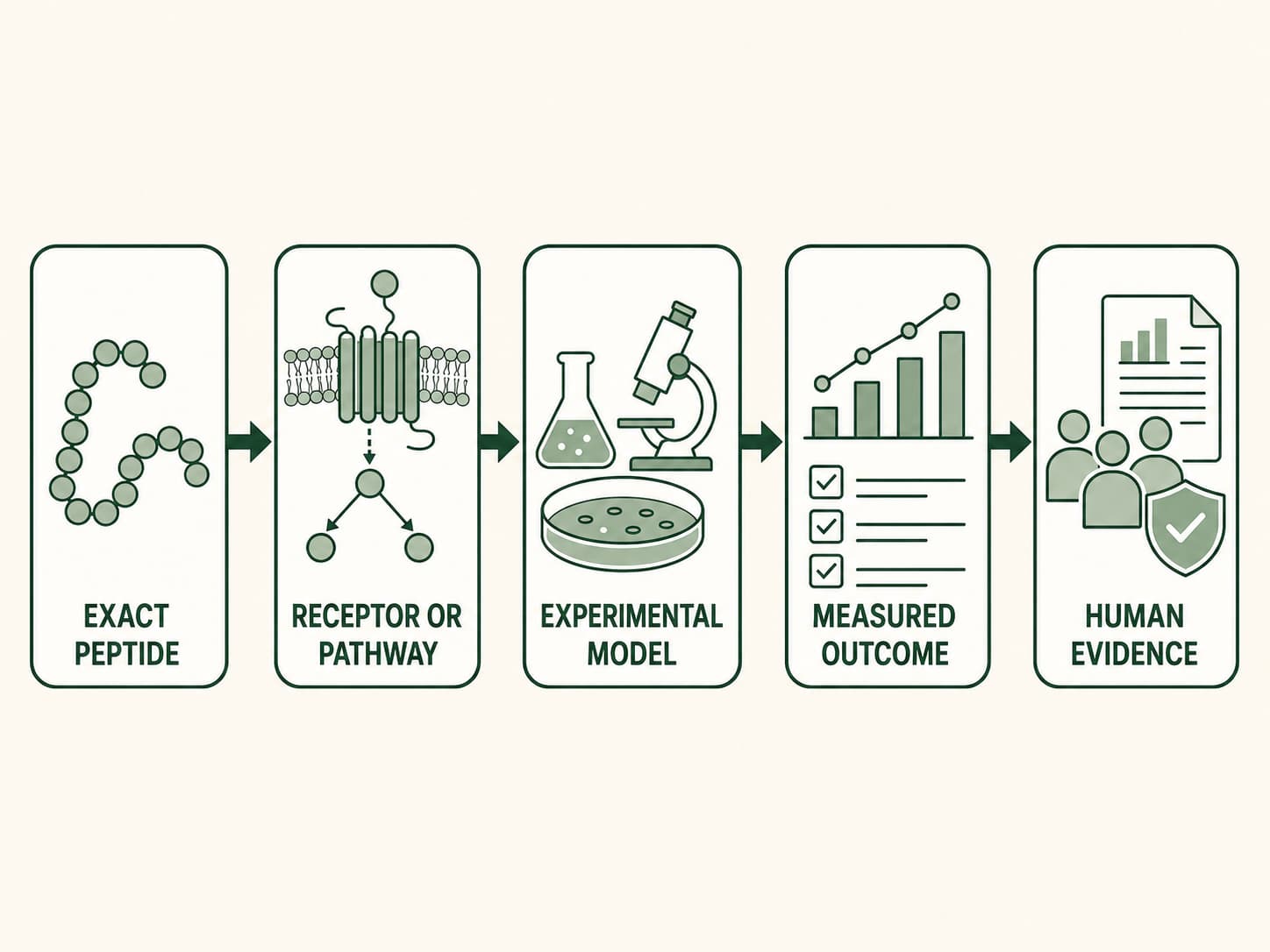
Figure 3: From peptide mechanism to human evidence. Evaluating peptide research requires following the evidence from the exact peptide and its receptor or signalling pathway through the experimental model and measured outcome to supporting human evidence.
Skipping one of those steps can turn a valid mechanistic observation into an unsupported health claim.
For compound-specific safety questions, continue with Are All Peptides Safe? Why the Answer Depends on the Compound.
For the distinction between authorised medicines and experimental substances, read FDA-Approved Peptides vs Experimental Peptides.
What Peptide Signalling Ultimately Depends On
Peptide signalling is specific rather than universal.
A peptide has to reach a responsive cell, interact with a compatible receptor and activate the signalling machinery available in that cell. The receptor may initiate G-protein signalling, second-messenger production, phosphorylation or another intracellular pathway.
The most useful way to understand peptide biology is therefore to follow the complete sequence:
Peptide → receptor → signalling pathway → tissue → experimental model → measured outcome → human evidence
That framework explains why different peptides can produce very different biological responses and why the same peptide can behave differently across tissues.
It is also more informative than treating “peptides” as one category with one shared effect.
FAQs
What Is Peptide Signalling?
Peptide signalling is cellular communication in which a peptide interacts with a compatible receptor and changes activity inside the target cell. Depending on the receptor, the signal can involve G proteins, second messengers, phosphorylation or other intracellular processes.
How Do Peptides Communicate With Cells?
Signalling peptides communicate with responsive cells by interacting with receptors. Receptor activation then starts intracellular signalling that can influence enzymes, ion movement, secretion, metabolism or gene activity.
Are Peptide Signalling And Peptide Hormones The Same Thing?
No. A peptide hormone is a type of signalling molecule. Peptide signalling describes the biological process through which peptide messages are recognised and converted into cellular responses.
Do All Peptide Receptors Use GPCR Signalling?
No. The GLP-1 receptor is a GPCR whose primary transduction mechanism includes Gs and adenylyl cyclase, whereas the insulin receptor is a receptor tyrosine kinase.
What Is A Second Messenger In Peptide Signalling?
A second messenger is an intracellular signalling molecule whose amount or activity changes after receptor activation. cAMP, IP3 and calcium ions are common examples in peptide-responsive signalling systems.
What Happens After A Peptide Binds To A Receptor?
The receptor changes its activity or conformation and begins transmitting the signal inside the cell. Depending on receptor type, this can activate G proteins, enzymes, second messengers, phosphorylation pathways or other signalling components.
Why Can The Same Peptide Affect Tissues Differently?
Different tissues can express different receptor subtypes and different amounts of those receptors. Their intracellular signalling machinery also varies, so the same extracellular signal does not necessarily produce an identical response in every cell.
How Does The Body Stop Peptide Signalling?
Signals can be limited through peptide degradation, second-messenger removal, receptor desensitisation, phosphorylation, internalisation, recycling and other feedback mechanisms. Research on GLP-1R and GIPR shows that the exact regulatory mechanism can differ between receptors.
Does Receptor Binding Prove That A Peptide Works In Humans?
No. Receptor binding or pathway activation demonstrates a molecular interaction under specific conditions. Human effectiveness and safety require appropriate clinical evidence; mechanistic findings alone cannot establish a clinical benefit.
Related reading
Peptide science
What Are Peptides? A Beginner’s Guide to How They Work
What are peptides? Learn what peptides are, how amino acid chains form peptides, what peptides do in the body, how they differ from proteins, and why their safety depends on the compound.
Peptide science
Peptides vs Proteins: What Is the Difference?
Learn how peptides and proteins differ in amino acid chain length, 3D folding, function, digestion and absorption, with examples including insulin and collagen.
Peptide science
Natural, Synthetic and Therapeutic Peptides Explained
Learn how natural, synthetic and therapeutic peptides differ, how peptides are produced, where the categories overlap, and what peptide type really tells you.